Learning Outline
Cell Structure & Function
Cell Structure
The generalized [eukaryotic] cell 
Plasma membrane
Cytoplasm – cell stuff
- Cytosol – cell solutions
- Organelles – cell structures
- Membranous and nonmembranous
Nucleus
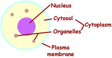
The “main” parts of a typical cell. ![]()
Cellular membranes (incl. plasma membrane)
Fluid mosaic model
- Phospholipid bilayer with embedded proteins and hybrid molecules

- Integral membrane proteins (IMPs) found within the phospholipid bilayer perform various specific functions
- Cholesterol (among phospholipid tails) stabilizes membrane
- Rafts
- Linked groups of membrane molecules that travel together like a raft within the fluid bilayer
- Linked groups of membrane molecules that travel together like a raft within the fluid bilayer
Functions — see table in Chapter 3 ![]()
- Cell membrane functions are cell functions (that is, many functions of cells that we will discuss are in reality jobs performed by the membranes of cells)

A “typical” cell model.
Click here for a larger image.
Campare to another cell model.
Other important basic cell structures
Nucleus
- Nuclear envelope
- Nuclear pores
- Nuclear pore complex (NPC) is the specific structure at each opening in the nuclear envelope
- Each NPC acts as a gatekeeper and transporter mechanism that regulates movement of molecules into and out of the nucleus
- Nuclear pore complex (NPC) is the specific structure at each opening in the nuclear envelope
- Nuclear pores
- Nucleoplasm
- Chromatin
- DNA plus protein
- Chromosome = condensed chromatin
- Primary genetic code of the cell
- Click here to see a slick view of DNA that will help you understand where it is and what it is
- Nucleolus — forms ribosome parts (rRNA)
- Chromatin
Mitochondrion (pl. mitochondria)
- Double membrane — inner membrane folded into cristae
- Interior is called matrix
- Involved in transfer of energy from fuel molecules to ATP
- Called the cell’s “power plant” or “battery charger”
- Serial Endosymbiosis Theory — SET (Lynn Margulis)
Ribosome
- Assembled as subunits of rRNA/protein in nucleolus
- Attach to mRNA strands (containing a gene) to guide assembly of amino acids into a polypeptide or protein
- Amino acids are brought to the ribosome by tRNA
|
Selected examples of important nucleic acids
|
|
| rRNA ribosomal RNA |
Forms ribosomes |
| mRNA messenger RNA |
Unfolded strand contains gene (code for one polypeptide); temporarily folds when leaving nucleus |
| tRNA transfer RNA |
Brings specific amino acids to ribosome and places them according to code on mRNA |
| nuclear DNA | “Master” genetic code in the nucleus |
| mDNA or mtDNA mitochondrial DNA | Additional “master” genetic code in the mitochondrion |
Endoplasmic reticulum (ER)
- Network of membranous canals and sacs
- ER extends outward from the outer boundary of the nucleus
- ER extends outward from the outer boundary of the nucleus
- Rough ER (RER) has temporarily-attached ribosomes
- Receives and processes polypeptides/proteins dropped off by ribosomes
- Also called granular ER
- Smooth ER (SER) has no ribosomes
- Also processes proteins and is site of enzyme action, including manufacture of membrane components (thus, it makes “new” membrane for the cell)
- Transports calcium ions (Ca++) into ER sacs, removing it from the cytosol (discussed later in course)
- Also called agranular or nongranular ER
Golgi apparatus
- Also called Golgi body or Golgi complex (named for Camillo Golgi) or dictyosome
- Stack of separate, flattened sacs
- Sacs made of membrane are often called cisternae (sing. cisterna)
- Processes, sorts, packages proteins sent by ER
Vesicles (literally “little vessels”)
- Fluid-filled “bubbles” of membrane
- Examples:
- Transport vesicles (such as ER or Golgi vesicles)
- Secretory vesicles
- Lysosomes contain lysing (digesting) enzymes
- Peroxisomes (bud from the ER) have enzymes that process H2O2 as they digest fats and detoxify poisons
Proteasome
- Hollow, drumlike cylinder made up of protein subunits
- Found throughout cytoplasm
- Breaks apart abnormal / misfolded proteins or proteins that are no longer needed
- Small proteins called ubiquitins tag proteins for destruction by proteasomes
- Breaks proteins into small segments, which are later broken apart to individual amino acids (which in turn are recycled)
- Failure to dispose of misfolded proteins could result in buildup of harmful plaques

Proteasome
A protein (green) is shown moving from top to bottom through the hollow proteasome. Middle part is cut away to see where active enzymes cut the protein into small segments, which then move out ofthe bottom end.
(click image to enlarge)
Cytoskeleton
- Made up of microfilaments, intermediate filaments, microtubules
- Adsorption of water on proteins and cross-linking of proteins gives cytoplasm a gel consistency
- Adsorption of water on proteins and cross-linking of proteins gives cytoplasm a gel consistency
- Functions include support, movement of cell, movement within cell, cell shape, connections with other cells, sensation
- Centrosome = microtubule-organizing center (MTOC)
- Includes two cylindrical centrioles
- Molecular motors
- Help to move materials or organelles within cell
- Provide power to move the cytoskeleton (and thus move or change the shape of cells
- Examples: dynein, myosin, kinesin
- Cell extensions
- Microvilli (sing. microvillus)
- Increase surface area for absorption
- Cilia (sing. cilium)
- Groups of moving cilia move fluids along the surface of a sheet of cells
- Primary cilium
- Most cells have one or more (blood cells do not)
- Have a sensory function (taste, smell, movement, etc)
- Involved in replication of centrioles
- Groups of moving cilia move fluids along the surface of a sheet of cells
- Flagella (sing. flagellum)
- Only the sperm cell has one
- Molecular motors at base allow flagellem to move, thus enabling swimming of sperm
- Microvilli (sing. microvillus)
Extracellular matrix (ECM)
- Material outside of cells
- The ECM is a complex arrangement of fibers and other molecules that interact with cells to perform body functions
- See the discussion in Chapter 5 of Anatomy & Physiology textbook
Cell connections
- Cells must be held together in a multicellular organism, or the tissues would simply fall apart
- In some tissues, cells are held together by fibrous “nets” that are not part of the cells themselves
- In some tissues, cells form junctions with each other

Desmosomes
- Spot desmosomes: small patches of filaments from adjoining cells “tangle” together like Velcro patches, holding cells together (example: skin cells)

Spot desmosome.
click to enlarge
-
- Belt desmosomes: connecting band (rather than small patch) encircling the cell and connecting it to nearby cells
- Belt desmosomes: connecting band (rather than small patch) encircling the cell and connecting it to nearby cells
- Tight junctions
- Bands of protein units in adjoining cells “snap together” to form a tight seal all the way around one “end” of a cell, forming a sort of “collar” that sticks to the collars of nearby cells and thus forms a seal to prevent molecules from passing by a membrane made of cells held together by tight junctions (example: lining of intestines)
- Bands of protein units in adjoining cells “snap together” to form a tight seal all the way around one “end” of a cell, forming a sort of “collar” that sticks to the collars of nearby cells and thus forms a seal to prevent molecules from passing by a membrane made of cells held together by tight junctions (example: lining of intestines)

Tight junction.
click to enlarge
- Gap junction
- Protein units form channels that link together to form “tunnels” that lead from one cell to the next
- This arrangement not only joins cells structurally but also functionally, because molecules can move back and forth through gaps and the plasma membrane of each cell is now a continuous sheet—as if it’s now one giant cell (example: heart muscle cells)

Gap junction.
click to enlarge
![]() Explore cell structure by clicking the icon . . .
Explore cell structure by clicking the icon . . . ![]()
For an interesting alternate hypothesis about basic cell structure, I highly recommend the book Cells, Gels, and the Engines of Life by Gerald Pollack
Cell Function
Transport concepts
- Passive forms of transport — do not require cell expenditure of energy
- Particles move down their concentration gradient
- That is, particles move from area of high concentration to area of low concentration
- If possible, particles eventually reach a dynamic equilibrium in which there is no difference in concentration
- Equilibration = process of reaching an equilibrium
- Particles move down their concentration gradient
|
Terms related to diffusion
|
|---|
| Solution — liquid mixture, usually composed of a liquid solvent and one or more dissolved particles (solutes)
Solute — a dissolved particle Solvent — a liquid into which other particles may dissolve <EXAMPLE: seawater is a solution in which sea salts are the solutes dissolved in the solvent water> Permeable — describes a structure through which substances may move; impermeable means that the structure does not permit a substance to pass through Permeant — describes a substance that is able to move through a structure; impermeant means that the substance cannot pass through Semi-permeable — describes a structure through which some, but not all, substance may pass Selectively permeable — describes a living structure that is able to choose which (and when) particular substances may move through it Conductance — the ease with which a substance may pass through a structure < EXAMPLES: A cell membrane may be permeable to oxygen but not to sodium ions, thus we say that the membrane is impermeable to sodium and that sodium is therefore an impermeant solute. Oxygen is a permeant solute. However, the cell may construct sodium channels and choose to open them under certain conditions. Thus we say that this membrane is selectively permeable to sodium. When the sodium channels open to allow sodium ions to pass through (be conducted through), we say that sodium conductance has increased. The more sodium channels open, the greater the sodium conductance. > |
|
Semipermeable membrane. |

-
- Simple diffusion
- Particles pass through cell membrane as they diffuse down their concentration gradient
- Depends on how membrane-soluble the particles are
- Particles pass through cell membrane as they diffuse down their concentration gradient
- Mediated transport (not-so-simple diffusion)
- Transporters are required
- Cellular membrane structures that serve as gateways to permit movement of particles through the membrane
- The shape & size of transporters determine what can pass through; if the shape changes, the ability to transport may change
- Channel-mediated passive transport
- Particles diffuse through membrane channels
- Some channels are gated (that is, they can open and close to selectively regulate conductance)
- Particles diffuse through membrane channels
- Carrier-mediated passive transport
- Particles diffuse through carrier mechanisms in a membrane
- Also called facilitated diffusion
- Transporters are required
- Osmosis — diffusion of water in presence of impermeant solutes
- Aquaporins — water channels that facilitate osmosis
- Osmotic pressure (actual vs. potential)
- Pressure that develops (actual) or could develop (potential) when water moves (osmoses) across a membrane and changes the volumes (and thus, the pressures) on both sides of the membrane.
- Isotonic — solution with same potential osmotic pressure as cell
- There is no net water movement into or out of cell
- There is no net water movement into or out of cell
- Hypertonic — solution with higher potential osmotic pressure than cell
- There is net movement of water INTO a hypertonic solution (out of cell)
- Hypotonic — solution with lower potential osmotic pressure than cell
- There is net movement of water OUT OF a hypotonic solution (into cell)
- Aquaporins — water channels that facilitate osmosis
- Simple diffusion

Red blood cells in solutions with different osmotic pressures.
The labels at the top describe the solution and not the intracelluar fluid.
Active forms of transport — require cell expenditure of energy
- Ion pumps (“active transport”)
- Particles are moved up their concentration gradient
- Pumps are carriers that use energy transferred from ATP
- Cotransport
- Particles are moved in the same direction by the same mechanism
- Also called symport
- Countertransport
- Particles are moved in opposite directions by the same mechanism
- Also called counterport or antiport
- Bulk transport by vesicles
- Exocytosis
- Moves large number of molecules OUT OF a cell
- Internal vesicle moves to plasma membrane and “pops open” releasing material from vesicle
- Endocytosis
- Moves large number of molecules INTO cell
- Plasma membrane pinches in, trapping extracellular material into a vesicle
- Two types:
- Phagocytosis
- Chunks are brought into cell (literally, “cell eating”)
- Pinocytosis
- Fluids are brought into cell (literally, “cell drinking”)
- Phagocytosis
- Exocytosis
Cell Life Cycle
All organisms have “life cycles” of development and reproduction—so do cells ![]()
- Parent (mother) cell divides to produce two genetically identical daughter cells
- Daughter cells may not be the same size or have exactly equal number of organelles
Phases ![]()
![]()
- Interphase (in-between phase)
- Phase during which cell is not actively dividing
- G1 phase (first growth [gap] phase) — new daughter cell is growing
- S phase (synthesis phase) — cell prepares for eventual cell division by replicating nuclear DNA
- So that there are now two identical sets (so that each daughter cell will receive one complete, identical set)
- Each DNA molecule splits (unzips) and new, complementary nucleotides fill in the missing side
- Each daughter DNA molecule (chromatid) is held together at the centromere
- G2 phase (second growth [gap] phase) — cell continues to grow, often “stockpiling” extra cytoplasm for an eventual split
- Centrioles (in the centrosome) are replicated during interphase
- Mitosis
- Coordinated division of the nuclear DNA and equal distribution to each daughter cell
- Provides genetic integrity
- Also called nuclear division or M phase
- Phases of mitosis
- Prophase (preliminary phase)
- Nuclear envelope dissolves
- Chromatin (DNA) strands shorten into compact chromosomes (each chromosome is made up of two chromatids held together by a centromere)
- Each centriole moves toward an opposite pole of the cell, constructing a spindle of fibers (microtubules) across the cell
- Prophase (preliminary phase)
- Coordinated division of the nuclear DNA and equal distribution to each daughter cell

![]()

Prophase
-
- Metaphase (positioning phase) — chromosomes align at equator of cell and attach to spindle fibers

Metaphase
-
- Anaphase (separation phase) — spindle fibers pull toward poles, separating the chromatids to form individual chromosomes moving away from their sisters

Anaphase
-
- Telophase (end phase) — everything goes back to the way it “should be”—the chromosomes unwind to chromatin, the nuclear envelope reforms, the spindle is dismantled

Telophase
- Cytokinesis — the “pinching in” of the membrane and eventual separation of the two daughter cells; not as precise as nuclear division (mitosis); overlaps the end of mitosis

Cytoknesis
- Daughter cells are now separate and in G1 of interphase
Chromosome numbers
Diploid number = 46 (for humans)
Haploid number = 23 (1/2 of diploid number)
Daughter cells should always have the diploid number of chromosomes—except egg cells and sperm cells, which should have the haploid number.
Differentiation of cells
- Different cell lines develop (differentiate) to specialize in different functions
- Stem cells are the “generic” ancestor cells
- Apoptosis — programmed cell death
- Allows the body to clear out older cells and make room for newer cells
Control of cell reproduction
- Neoplasms = tumors
- Benign tumor (nonspreading) or malignant (spreading = metastasis)
- Hyperplasia = too many cells
- Anaplasia = undifferentiated cells
- Dysplasia = disorganized tissue with abnormal cells
Cellular metabolism
- Requires enzymes
- Anabolism (building larger molecules)
- Example: protein synthesis
- Catabolism (breaking down large molecules)
- Covered in more detail later (for example, when we discuss muscle function)
![]() Optional: Click here for another helpful outline Getting Energy from the Pre-A&P course.
Optional: Click here for another helpful outline Getting Energy from the Pre-A&P course.
This is a Learning Outline page.
Did you notice the EXTRA menu bar at the top of each Learning Outline page with extra helps?
Last updated: November 16, 2019 at 0:08 am