This is a brief review of the concept of tissues in the context of human biology. You will not be expected to be able to identify them by sight in the onlines tests or exam. That will come later, in your A&P lab course.
Four main types of tissues: epithelial, connective, muscle, nervous
Extracellular matrix (ECM)
Complex, nonliving material filling the spaces between cells
Some tissues (epithelial) has little ECM and a lot of cells; some tissues (connective) have a lot of of ECM and few cells
Made up of
Water
Proteins
Glycoproteins – part carbohydrate, part protein
Proteoglycans – protein backbone with carbohydrates attached
Functions
Help hold tissues together
Communication between ECM and cell or among cells
Extracellular matrix
Extracellular matrix (ECM)
Immunofluorescent staining of fibroblast cells ( their nuclei are greenish here) reveals the extracellular matrix (bluish and turquoise areas)
Epithelial tissues
Function
Cover and line (form membranes)
Secrete (form glands) – “secretory tissue”
Endocrine glands (ductless glands) secrete hormones into blood
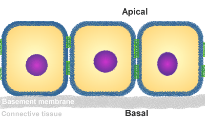
Desmosomes – “tangled filaments” hold cells together as in Velcro fasteners
Gap junctions – channels in adjacent plasma membranes form “tunnels” that hold cells together structurally and functionally
Syncytium – group of cells held together by gap junctions and acting (in some ways) as a single, giant cell
These occur mainly in cardiac muscle and nervous tissue—but are discussed here for the sake of convenience
Tight junctions – rows of connecting proteins, like “snaps” on a jacket form a collar-like seal all the way around a cell (as the plastic yoke on a six-pack)
tight junction
Basal surface of each cell is anchored; apical surface of each cell is is “free”
Basal layer attached to underlying connective tissue by glue-like basement membrane
Epithelial types are named for
Number of layers
Simple = one layer of cells
Stratified = more than one layer of cells (from stratum = layer)
Pseudostratified = looks like more than one layer, but because all cells touch the basement membrane it’s really just one layer
Shape of cells in outermost (“free surface”) layer (as seen in cross-section)
Squamous = flat, scale-like cells
Cuboidal = about as tall as wide
Columnar = taller than wide
Transitional = shape depends on how stretched the membrane is
Examples of epithelial tissue types
Simple squamous
Simple cuboidal
Simple columnar
Pseudostratified columnar
Stratified squamous
Keratinized (has outer layers of dead cells that have filled with tough, waterproof protein called keratin—as in skin)
Nonkeratinized
Transitional (stratified
epithelial categories
Connective tissues
Function
Connects body parts in any of several ways
Many different functions, really
Structure
Dominated by extracellular material (“extracellular matrix” or ECM)—with few, scattered cells
Matrix contains fibers (made by fibroblast cells) and other materials
Elastic fibers – made of elastin and stretch easily, then recoil (also called yellow fibers)
Collagen fibers – made of collagen and do not stretch (much) but are strong and flexible (also called white fibers or collagenous fibers)
Major types of connective tissues
Fibrous connective tissues
There are several ways to classify fibrous connective tissues—most commonly, they are categorized by the structure of the matrix (type and arrangement of fibers)
Loose (areolar) connective tissue has collagen and elastin fibers scattered loosely
Adipose tissue forms when fat-storing cells in areolar tissue enlarge as they accumulate more [triglyceride] fat
Dense fibrous connective tissue has a dense arrangement of collagen fiber bundles
Regular – has rougly parallel bundles of collagen fibers
Irregular – has hodgepodge, irregular arrangement of collagen fibers
Reticular tissue is a netlike meshwork of fine collagen fibers that helps hold tissues in place and sometimes helps to filter particles from fluids passing through it
3 connective types
Three types of fibrous connective tissue.
The third image is regular dense fibrous connective tissue.
Cartilage
Chrondrocytes make cartilage matrix
Found within spaces called lacunae (sing. lacuna = “lake”) giving cartilage a “Swiss cheese” appearance
Hyaline cartilage – some collagen in matrix
Elastic cartilage – some collagen and some elastin in matrix
Fibrocartilage – dense collagen in matrix
swiss cheese
Swiss cheese.
A model for cartilage. Compare this image to that of hyaline cartilage (below).
Bone
Osteocytes (within lacunae) surrounded by collagen fibers encrusted with calcium salts
Spongy bone – irregular beams of bone surrounded by red bone marrow (soft, blood-forming tissue)
Compact bone – denser type of bone made up of cylindrical units composed of concentric layers (lamellae) of bone matrix
Blood
Liquid matrix (blood plasma) and circulating blood cells
Blood cells are also called “formed elements” because plasma is “unformed” (taking the shape of its container)
RBCs = red blood cells, WBCs = white blood cells, platelets = thrombocytes
blood, bone, and cartilage
Blood, bone and cartilage
Bone is sometimes called “osseous” which means “bony” (compact bone is shown)
Muscle tissue
Function
Contraction
May be “voluntary” or “involuntary”
Structure
Cylindrical cells or “muscle fibers”
Muscle fibers have highly organized cytoskeleton that “slides together” like a split deck of cards to contract the fiber
Three types: skeletal, smooth, cardiac
Skeletal muscle
Also called “striated” because of striped appearance of overlapping filaments of cytoskeleton
Voluntary muscle
Connected to skeleton
Cardiac muscle
Faintly striated
Involuntary
In heart wall
Branched fibers held together end-to-end by gap junctions, forming intercalated disks that connect cardiac fibers into a network
Forms a syncytium, so that a region of the heart wall will contract at one time, as if one giant cell
Smooth muscle
Not striated (cytoskeleton organized differently)
Involuntary
In walls of hollow organs (except heart)
3 types of muscle tissue
Muscle tissue types.
Nervous tissue
Neurons
Conducting cells
Connected at synapses that act as switches, allowing information storage and processing
Glia (also called neuroglia)
Support cells (functional and structural support)
Involved in modulating neuron function
brain tissue
Brain tissue.
Click the image to see a larger view, where a mixture of neurons and glia can be seen.
This is a Learning Outline page.
Did you notice the EXTRA menu bar at the top of each Learning Outline page with extra helps?